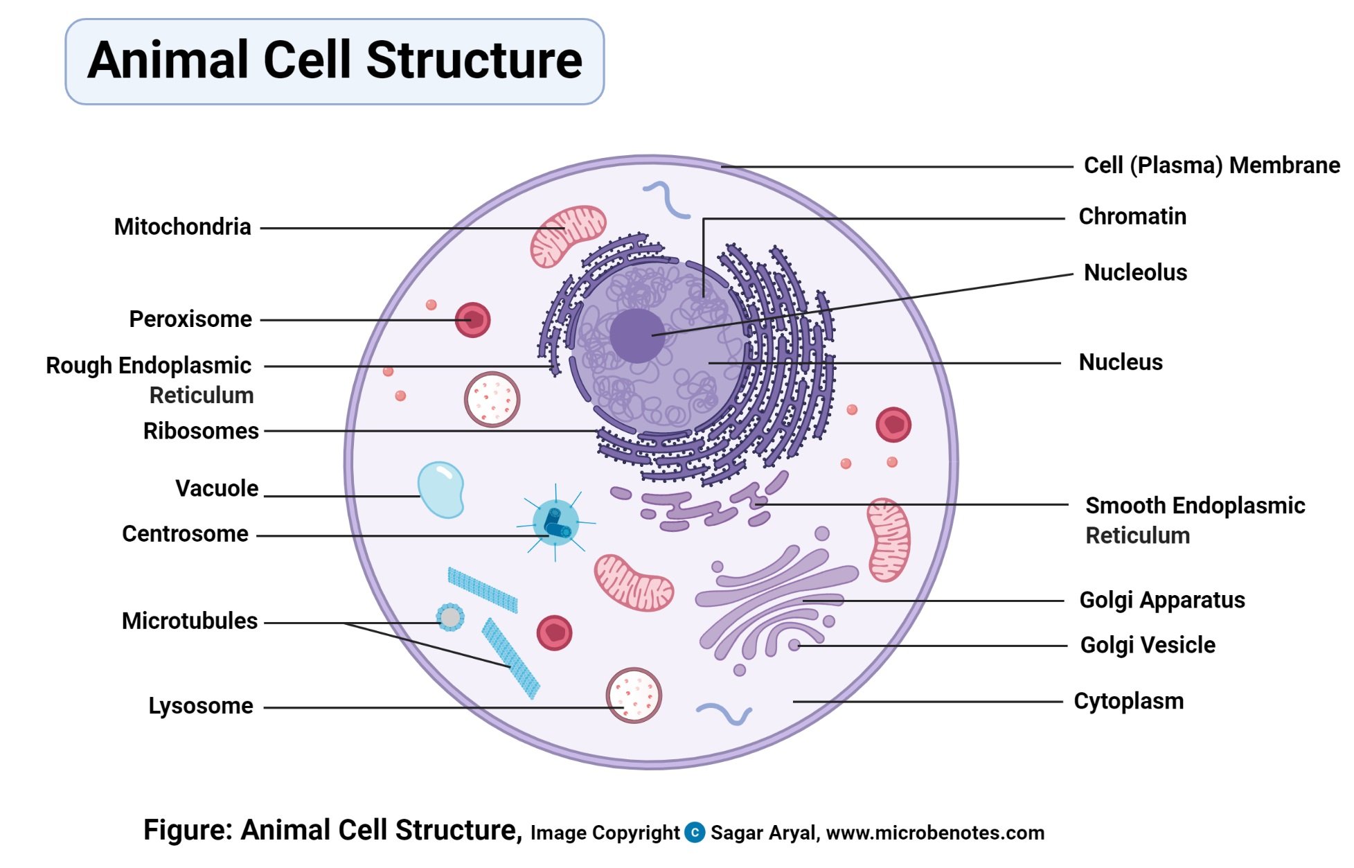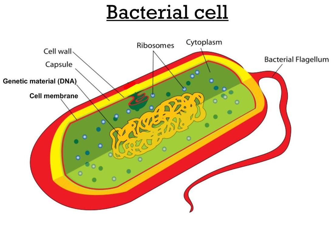

#Cell diagram labeled free
(In eucaryotic plasma membranes, larger tears are repaired by the fusion of intracellular vesicles.) The prohibition against free edges has a profound consequence: the only way for a bilayer to avoid having edges is by closing in on itself and forming a sealed compartment ( Figure 10-5). A small tear in the bilayer creates a free edge with water because this is energetically unfavorable, the lipids spontaneously rearrange to eliminate the free edge. The same forces that drive phospholipids to form bilayers also provide a self-healing property. In this energetically most-favorable arrangement, the hydrophilic heads face the water at each surface of the bilayer, and the hydrophobic tails are shielded from the water in the interior. (more.)īeing cylindrical, phospholipid molecules spontaneously form bilayers in aqueous environments. (B) A lipid micelle and a lipid bilayer seen in cross section. (A) Wedge-shaped lipid molecules (above) form micelles, whereas cylinder-shaped phospholipid molecules (below) form bilayers. Packing arrangements of lipid molecules in an aqueous environment. This free energy cost is minimized, however, if the hydrophobic molecules (or the hydrophobic portions of amphipathic molecules) cluster together so that the smallest number of water molecules is affected. Because these cage structures are more ordered than the surrounding water, their formation increases the free energy. If dispersed in water, they force the adjacent water molecules to reorganize into icelike cages that surround the hydrophobic molecule ( Figure 10-3). Hydrophobic molecules, by contrast, are insoluble in water because all, or almost all, of their atoms are uncharged and nonpolar and therefore cannot form energetically favorable interactions with water molecules. As discussed in Chapter 2, hydrophilic molecules dissolve readily in water because they contain charged groups or uncharged polar groups that can form either favorable electrostatic interactions or hydrogen bonds with water molecules. It is the shape and amphipathic nature of the lipid molecules that cause them to form bilayers spontaneously in aqueous environments.

The kink resulting from the cis-double bond is exaggerated for emphasis. This example is phosphatidylcholine, represented (A) schematically, (B) by a formula, (C) as a space-filling model, and (D) as a symbol. Differences in the length and saturation of the fatty acid tails are important because they influence the ability of phospholipid molecules to pack against one another, thereby affecting the fluidity of the membrane (discussed below). As shown in Figure 10-2, each double bond creates a small kink in the tail.

One tail usually has one or more cis-double bonds (i.e., it is unsaturated), while the other tail does not (i.e., it is saturated). The tails are usually fatty acids, and they can differ in length (they normally contain between 14 and 24 carbon atoms). These have a polar head group and two hydrophobic hydrocarbon tails. The most abundant membrane lipids are the phospholipids. All of the lipid molecules in cell membranes are amphipathic (or amphiphilic)-that is, they have a hydrophilic (“water-loving”) or polar end and a hydrophobic (“water-fearing”) or nonpolar end. There are approximately 5 × 10 6 lipid molecules in a 1 μm × 1 μm area of lipid bilayer, or about 10 9 lipid molecules in the plasma membrane of a small animal cell. Lipid-that is, fatty-molecules constitute about 50% of the mass of most animal cell membranes, nearly all of the remainder being protein. Membrane Lipids Are Amphipathic Molecules, Most of which Spontaneously Form Bilayers


 0 kommentar(er)
